

Outline
This half-body simulator is designed to simulate the real-world environment of ultrasound diagnosis in the human body, providing highly realistic and effective training conditions. It enables users to diagnose multiple lesions by simulating internal anatomical structures, such as bones, organs, and blood vessels. Through this immersive training, users can practice ultrasound examination techniques in a safe and true-to-life scenario, ultimately enhancing their practical skills and diagnostic accuracy.
Skills Gained
• Familiarity with human anatomical structures
• Interpreting ultrasound images
• Identifying pathologies via ultrasound
• Handling ultrasound machine & selecting appropriate probes
• Various centesis
• Ultrasound-guided liver and kidney puncture biopsy
Features
• Anatomy
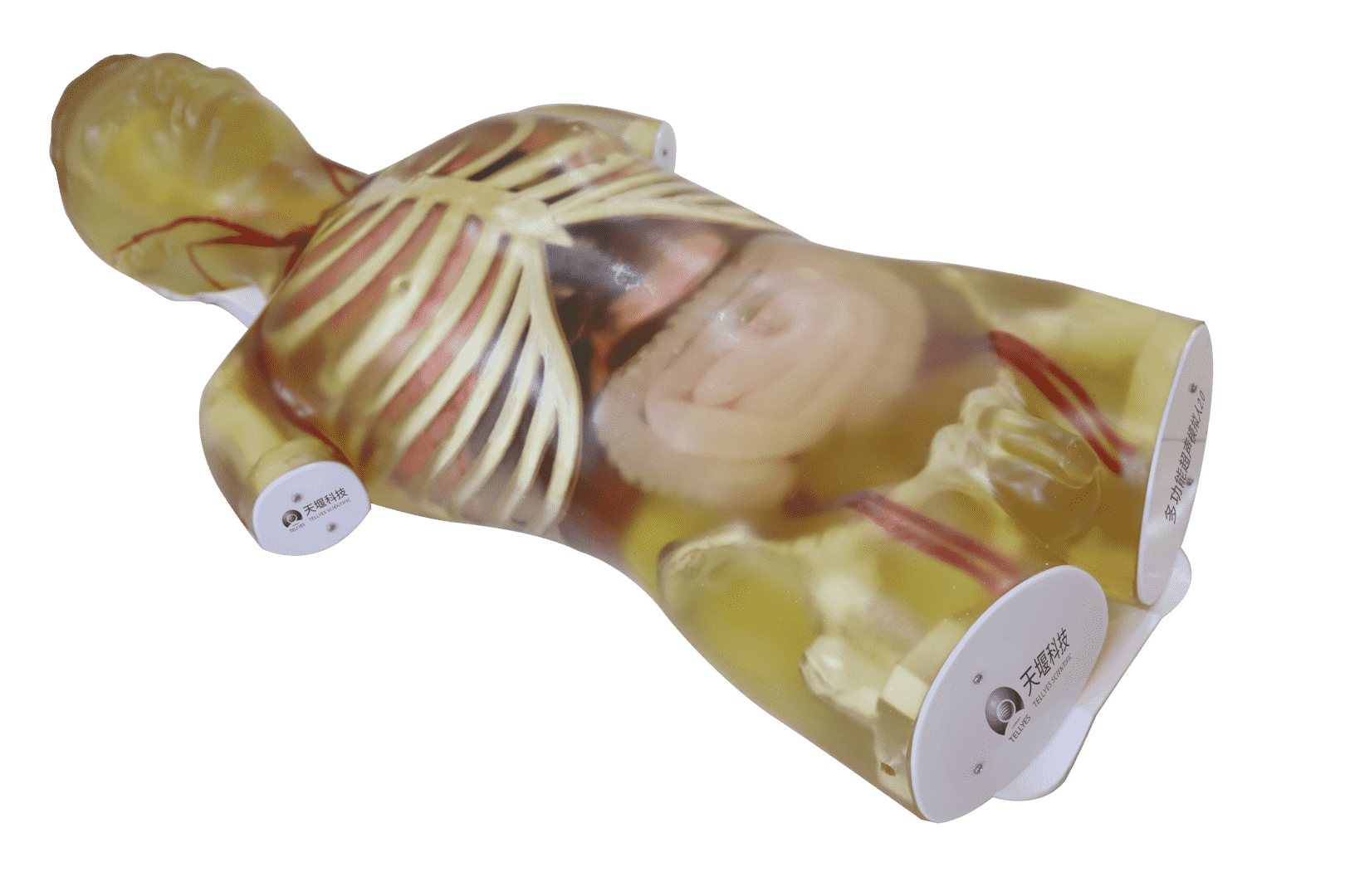
1) This half-body male manikin features a complete structure of the head, torso, organs and vessels.
2) Bionic skeletal structure includes ribs, sternum, sternal manubrium, clavicle, costal arch, xiphoid process, pelvis, femur. Accurate and palpable umbilicus.
4) Internal organs: lungs, heart, diaphragm, esophagus, stomach, duodenum, jejunum, large intestine, small intestine, liver, gallbladder, pancreas, spleen, kidneys, adrenal glands, ureters, and bladder.
• Realism
1) Compatible with clinical ultrasound machines to acquire lifelike diagnostic images during training.
2) The sonographic imaging demonstrates accurate organ sizing, true-to-life acoustic properties including organ dimensions, density, speed of sound propagation, attenuation, and echogenicity.
• Key features
Thoracic Region:
Heart:
• Intact heart structures including 4 chambers and valves.
• Allowing for simulating the standard subxiphoid 4-chamber view.
• Ultrasound images of 8 structures can be obtained, including the ventricles, atria, mitral valve, tricuspid valve, aortic valve, pulmonary valve, pulmonary artery, and aorta.
Lungs:
• True-to-life left and right lung structures, allowing for scanning by using an ultrasound probe in the long-axis orientation sequentially along the parasternal, midaxillary, posterior axillary lines, and the lateral scapular border. By scanning along the intercostal spaces in the short-axis orientation, the characteristic ultrasound image features of normal air-filled lungs with a visible pleural line can be obtained, and the ultrasound images of ribs and sternum can be clearly visualized.
Abdominal Region:
With vital organs, enabling the images of longitudinal section along the midline of the abdomen, as well as at the levels of the xiphoid process, umbilicus, and suprapubic region.
Liver:
1)The liver has 6 anatomical structures, including left lobe, right lobe, caudate lobe, ligamentum teres hepatis, first hepatic portal, second hepatic portal, etc., containing the portal venous system and hepatic veins, with an intact liver capsule
2)Lesions:
• Hepatic parenchyma was visualized in ultrasound examination. Sonography demonstrates hepatic space-occupying lesion.
• Space-occupying lesions: hyperechoic, hypoechoic, isoechoic, and anechoic cystic lesions.
• Lesion morphology: oval, round, and irregular. The irregular lesions appeared hypoechoic, contained calcific foci, and exhibited ill-defined margins.
3)Ultrasound-guided liver biopsy is supported.
4)The liver can be divided into 4 sectors via the left, right, and middle hepatic veins during the ultrasound examination. At the transverse plane, the horizontal sections of the right and left portal vein branches present as a linear or band-like transverse fissure structure, further subdividing the hepatic sectors into 8 segments according to the Couinaud Nomenclature.
Gallbladder
1) Gallbladder contains 7 anatomical structures, including cystic duct, common bile duct, left hepatic duct, right hepatic duct, cystic neck, cystic body, and cystic fundus.
2) Lesions: gallstones and polyps, during the ultrasound examination, gallstones appear as hyperechoic with posterior acoustic shadowing.
Kidneys
1) Both kidneys are anatomically intact and bean-shaped.
Bilateral renal regions can be examined via transverse and longitudinal plane ultrasound scanning in the left and right upper abdominal quadrants, as well as through dorsal and flank approaches, which allows for the visualization of 7 structures, including renal capsule, renal cortex, renal medulla (renal papilla, renal pyramid, renal column), renal pelvis, major calyces, minor calyces, and ureter.
2) Lesions:
• 2 renal calculi lesions at the major calyces and renal pelvis, presenting hyperechoic echogenicity with posterior acoustic shadowing.
• 1 renal tumor lesion at the upper pole of the left kidney, with indistinct margin, presenting hyperechoic echogenicity without posterior acoustic shadowing and internal echotexture is homogeneous.
• 1 renal tumor lesion at the upper pole of the right kidney, presenting isoechoic echogenicity with clear margin and internal echotexture is heterogeneous, containing hyperechoic calcified foci.
3) Ultrasound-guided renal biopsy is supported.
4) The adrenal glands can be detected superior to the bilateral renal areas.
Pancreas:
1) The pancreas contains the pancreatic duct. A well-defined margin with homogeneous internal echotexture from the pancreatic parenchyma and a slender pancreatic duct can be visualized at the upper abdomen.
2) Lesions: When scanning the pancreatic head, a solid hypoechoic mass with clear margin is visualized, consistent with characteristic imaging features of a pancreatic space-occupying lesion.
Spleen:
1)The spleen contains splenic hilum. The splenic capsule and splenic parenchyma can be visualized during ultrasonographic scanning along the anterior and posterior axillary lines.
2) Lesions:
• 1 splenic cyst lesion demonstrates a circular anechoic area with a smooth-walled capsule on ultrasonography.
• 1 splenic tumor that demonstrates an ovoid hyperechoic mass with clear margin.
Digestive system
The model incorporates 11 anatomical structures, including esophagus, cardia, gastric body, lesser curvature, greater curvature, gastric antrum, pylorus, duodenal bulb, descending duodenum, horizontal duodenum, and adjacent pancreatic, which exhibit characteristic contrast-enhanced ultrasound imaging features during ultrasound examination.
Built-in blood vessels:
All the arteries and veins are designed with true-to-life shape, vessel diameters, sizes, and anatomical course.
When pressure is applied with an ultrasound probe, venous vessels can be compressed flat, while arterial vessels maintain the shape. True-to-life vessel course and lumen diameter are visualized during the ultrasound examination, with simulated blood inside, which allows for the measurement of vessel length and internal diameter.
Cephalic and cervical region: jugular vein and carotid artery
Thoracic region: Supports ultrasound imaging of 9 vascular structures, including: aorta, aortic arch, superior vena cava, inferior vena cava, pulmonary artery, pulmonary vein, subclavian artery, subclavian vein, axillary artery, axillary vein
Abdominal region: Supports ultrasound imaging of 29 vascular structures, including:
• Inferior vena cava, abdominal aorta, celiac axis.
• Common hepatic artery, proper hepatic artery, portal vein, right intrahepatic portal vein branch, left intrahepatic portal vein branch, interlobar vein, hepatic vein, left hepatic vein, right hepatic vein, middle hepatic vein.
• Superior mesenteric artery, superior mesenteric vein, inferior mesenteric artery, inferior mesenteric vein.
• Splenic artery, splenic vein.
• Left renal artery, left renal vein, right renal artery, right renal vein.
• Left testicular artery, right testicular artery
• Common iliac artery, common iliac vein, external iliac artery, external iliac vein.
Stock code :833047
Address:2nd & 3rd Floor, West 6th Building, 18 West HaiTai Road, Tianjin, China
Postcode:300384
Phone:4006-355-510
+86-22-83711066
Fax:+86-22-83711065
Email:info@tellyes.com

 Ultrasound